Surgical Essentials: Scalpel Blades and Handles

Embed Block
Add an embed URL or code. Learn more

It’s been a while since we did a surgically-focused episode - we’ve previously done a series on laparoscopy and hysteroscopy, as well as on sutures and needles. Today, let’s focus in on an essential surgical instrument - the scalpel!

Additional reading: British Journal of Surgery Oct. 2022 review (also, the author Dr. Ron Barbosa is on Twitter and does some great surgical tweetorials!) 

History of the modern scalpel

  • Morgan Parker, a 22 year old engineer at the time, patented a locking scalpel handle and blade system in 1915 to replace what previously were often single-piece instruments without a replaceable blade.

    • His original design (slightly modified) is still what we use today!

  • Parker initially numbered handles 1-9 and blades 10-20; while this has been somewhat modified/expanded, the nomenclature largely remains the same.

    • We’ll talk about the most common handles and blade types today.

Scalpel handles

  • You’ve probably never had to ask for these in a surgical tray – so let’s review!

  • The number three handle is most commonly used in surgical specialties:

    • Flat shape

    • Some serrations near the blade attachment area to provide better grip for surgeon

    • Fits blade numbers 10-19

    • Modifications include the 3L (long-handle scalpel) and 3L angled (long-handle with a slight angulation).

  • The number four handle fits larger blades (#20 and above), but otherwise is very similar to the #3. 

  • The number seven handle is very narrow and meant for precise, fine work – not typically used in OB/GYN or subsepcialties – more common in head/neck/ENT, plastics, neurosurgery, and dentistry.

Barbosa, BJS, 10/2022

Scalpel blades

  • You may be more familiar with these, but likewise may not have had to ask for them before!

  • The number ten blade is used to make longer skin incisions for laparotomy, or for shorter cuts where a wide blade is ideal (i.e., hysterotomy). 

    • This is probably what you’re most familiar with in OB/GYN applications. 

    • You may also encounter a number 22 blade, which is essentially a larger version of the #10.

  • The number eleven blade is triangular, long, and has a sharp point with an edge on one side. 

    • Its shape is best suited for a stab incision - for instance, for laparoscopic port incisions, Bartholin’s gland cruciate incisions, and the like.

    • Its shape is not great though for excising anything - it’s really pointy!

  • The number fifteen has a small, curved cutting surface as well as a pointed tip.

    • You can use this for a stab incision at the point, and a more controlled incision for excising tissue with the curved portion.

    • Great for working in tight spaces versus a 10 blade for excision (i.e., oncology cases or urogyn cases – think about sharply cutting on your cardinal ligament bites – a 15 blade on a 3L handle is great for this!)

    • Also great for stab incisions, and many folks may prefer a 15 to an 11 blade for Bartholin’s or laparoscopy incisions.

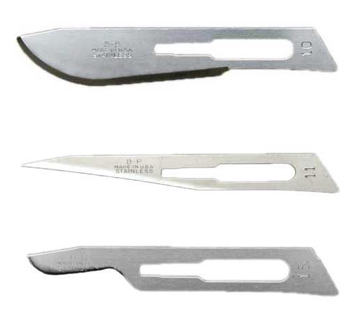
10 blade (top), 11 blade (middle), 15 blade (bottom)

Scalpel blade materials, and disposables versus regular blade/handles

  • Disposables are great and often very available for outpatient procedures or for emergencies

  • The blades between disposable and regular blades are the same shape/size/nomenclature, so there’s no difference in that regard.

    • However, the regular blades tend to be a bit thicker on the back, non-cutting surface of the blade, which gives a bit more structure and may feel sturdier when cutting.

  • In terms of materials, the vast majority of scalpels we use will be made of carbon steel or stainless steel.

    • Steel blades can also have other compositions or coatings that can help with retaining sharpness and/or resisting rusting/corrosion.

  • Other materials used in modern blades include ceramic, titanium, diamond, sapphire, and obsidian.

    • Many of these - especially ceramic and obsidian - are extremely sharp, and can be chosen because they are non-magnetic – so for MRI-guided procedures, they are preferred. However, they are so sharp that they can be very dangerous in poorly trained hands - so we wouldn’t use these unless you have a great reason to do so! 

How do I hold a scalpel?

  • Intern struggle – and the truth is that it depends!!! 

    • For larger blades - i.e., a #10 blade or a #20 or above - the best grip is a “palmar” or “violin grip,” in which you have your index finger atop the handle, and use your other fingers to hold the body of the handle, with the back part of the handle under your palm.

      • This allows for precision with the long, wide cuts you would typically make with this blade.

    • For smaller, pointier blades – a #11 or #15 - the “pencil grip” is preferred.

      • This allows for precision with your “stab” incision or for those tight/deeper spaces. 

Barbosa, BJS 10/22: Palmar/”Violin” grip, for larger blades (#10, #20 and above)

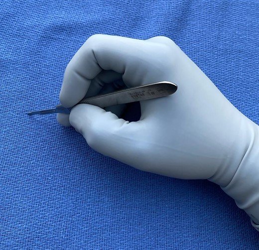
Barbosa, BJS 10/22: Pencil grip, for smaller blades (#11, #15)

Principles of Electrosurgery, feat. Dr. Gary Frishman

Here’s the RoshReview Question of the Week:

A 45-year-old woman in the postoperative recovery unit develops dyspnea. Her serum sodium is 130 mEq/L. Which of the following was the most likely distending medium used during her hysteroscopic monopolar fibroid resection?

Check your answer and get a sweet deal on RoshReview at the links above!


Today we welcome Dr. Gary Frishman to the podcast. He is a clinical professor of obstetrics and gynecology at the Warren Alpert Medical School of Brown University, and has served in varying leadership capacities at Brown as a residency program director and in other organizations, such as for MIGS Fellowship at ABOG, and within ASRM, SRS, and AAGL. While we were residents at Brown, Dr. Frishman used to share his wisdom with us on electrosurgery and we have wanted to put this podcast together for a long time! It’s more than just cut and coag!

What is electrosurgery?

  • Delivery of radio frequency waves that heat up tissue, which then causes tissue desiccation.

    • Cautery: not a specific term for this!

    • Electrosurgery represents alternating current where the body is part of the conductive circuit.

      • Modulating the delivery of this electricity is how we can get differing tissue effects.

Monopolar and Bipolar

  • Bipolar: movement of electricity from one point to another direct point.

    • i.e., bipolar forceps, LigaSure device (has two ends)

    • More precise delivery of energy, less smoke.

  • Monopolar: movement of electricity from one point (in dispersive fashion) to a return point.

    • i.e., the “grounding pad” when you are using a Bovie / electrosurgical instrument.

      • Energy still goes through body, to a second electrode and back to the machine!

Modifying the Delivery of Energy

  • Waveforms:

    • Think about energy delivered as a garden hose with a spray nozzle at the end.

      • The amount of energy delivered is your watts setting on the machine (i.e., 35W).

        • This you can adjust linearly — you can go up to 40W, or down to 30W, but it’s a linear adjustment — less efficient.

      • If you adjust your spigot at the end of the nozzle, that is another way to adjust your energy delivery. Like if you get a dispersed spray (sprays all over the wall) versus a fine spray (that gives you a directed, straight line of water).

        • Cut: the directed, fine, straight line of water spray

        • Coag: the dispersed spray that goes everywhere

          • The energy is still our original 35W, it’s just differently applied!

      • Importantly, the cut and coag settings also are different in the time energy is applied.

        • Cut: energy is constantly applied — so, for example, 100W of energy delivered over 100 seconds.

          • Clinically, this delivers good cutting, but poor hemostasis.

        • Coag: energy is applied in bursts, and it’s only on 6% of the time — so 100W of energy delivered over 6 seconds.

          • It’s an uncontrolled, massive burst!

          • Clinically, this delivers a lot of lateral spread, and you get good hemostasis, but not great cutting.

        • Blend curve: tries to get you both: excellent cutting, excellent hemostasis, little thermal damage.

  • Spot size:

    • You can also adjust the spot size of energy delivery by changing the tip of delivery instrument (i.e., Bovie)

      • By flattening the tip, you make the spot size larger — much larger surface area to deliver energy across.

      • By using the fine point of the tip, your spot size is much smaller — delivers energy to more concentrated area (i.e., needle tip electrodes).

Complications of Electrosurgery

  • Direct coupling

    • Accidentally touch something metal with your electrosurgery device

      • i.e., touching a retractor or a laparoscope, which is in turn touching something else

      • Good news — just like with the “spot size,” if you touch a retractor, it’s such a large surface area it’s unlikely to cause damage. However, with smaller instruments, this can modify injury and cause injury.

        • Plastic trocars help prevent this in laparoscopic surgery.

  • Insulation failure

    • Tiny cracks in an instrument that can cause insulation to fail — as you might get inadvertent direct coupling from the device if electricity is leaking out through the insulation.

      • Fortunately this is very uncommon.

      • If there are gross breaks — don’t use the instrument.

      • Limit the use of coag (i.e., use cut exclusively) to prevent this kind of injury.

  • Capacitive coupling

    • Two conductors separated by an insulator

      • Energy is stored in the separated conductor, and can then deliver energy.

        • Less likely to occur with cut than coag

        • Jewelery and electrosurgery - a possible (though rare) complication of wearing jewelry in surgery.

Role of Tissue Resistance

  • Electricity heats up water in the cell

    • Cut: heats up water in the cell very rapidly, and it explodes.

    • Coag: heats up water in cell more slowly, and energy dissipates laterally.

  • Electricity follows path of least resistance

    • As tissue dessicates (water removed), it becomes harder for electricity to pass through.

    • Think about a car on cruise control, set to a speed of 30W:

      • If you’re going uphill, you’re going slower unless car adjusts — higher tissue resistance.

      • If you’re going downhill, you’re going faster unless car adjusts — lower tissue resistance.

  • Newer generators measure tissue resistance and can adjust your energy appropriately!

    • Newest bipolar instruments also take surgeon out of equation entirely — and automatically adjust energy to resistance and shut off automatically when resistance is so high energy can’t pass.'

  • Importantly, if you’re using monopolar — electricity will go around high-resistance areas! This is how you can get capacitive coupling to jewelry and other areas.

    • A “grounding pad” is usually very large to help accommodate for this and prevent this injury.

      • It’s also in two halves, and both must be connected to make the machine work.

      • Bipolar instruments reduce this risk significantly.

Fulguration

  • Taking the tip of the device and placing it close to, but not on the tissue — may have heard of this as “arcing” the device.

    • Use coag on this because you want a huge burst of energy to leap across the space.

    • This energy “follows the blood” back to the original bleeder.

Cutting on Skin??

  • You can! But you need to know how to deliver energy — need a small spot size and to use cut.

Hysteroscopy II: Complications and Troubleshooting

Here’s the RoshReview Question of the Week!

A 45-year-old woman in the postoperative recovery unit develops dyspnea. Her serum sodium is 130 mEq/L. Which of the following was the most likely distending medium used during her hysteroscopic monopolar fibroid resection?

Check out the answer and enter the QE QBank Giveaway at the links above!


Why do we do hysteroscopy?

  • Diagnostic

    • AUB

    • Infertility

    • Structural anomalies

  • Operative

    • IUD removal

    • Polyps

    • Fibroids

    • Septums

    • Intrauterine adhesions and Ashermans

    • Endometrial ablation

    • C/section scar (isthmocele) excision

    • C/section scar or cervical ectopics

    • Tubal cannulation

Complications of Hysteroscopy -check out ACOG CO 800!

  • Perforation

    • Most common complication - range 0.12-1.61%

    • Risk factors

      • Blind insertion of instruments

      • Cervical stenosis

      • Anatomic distortion - fibroids, adhesions, myometrial thinning, extreme anteversion or retroversion)

    • High index of suspicion

    • Decrease risk by using ultrasound or laparoscopic guidance

    • With dilation or with scope (unlikely)

      • Low risk of subsequent complications

    • During instrumentation

      • Increased risk of injury to extra-uterine structures

      • Requires evaluation - laparoscopy +/- laparotomy 

  • Fluid Overload

    • Rare - 0.2%

    • Risk factors - resection of large or deep lesions, high pressure setting

    • As reviewed in prior episode

      • Limit fluid deficit to 1000mL for electrolyte-free hypotonic media, 2500mL for electrolyte-rich isotonic media

      • Use fluid management systems, Designated individual to monitor fluid deficit, decreased deficit limits for patients with comorbidities

      • Other preventive measures

        • GnRH agonists (pre-op)

        • Intracervical vasopressin injection

        • Planning for staged procedures

    • Management

      • Stop procedure

      • Assess hemodynamic, neurologic, respiratory, CV status

      • Check labs - serum electrolytes, osmolality

      • Consider loop diuretic (Furosemide)

      • Consider hypertonic saline

  • Hemorrhage

    • 0.03-0.61%

    • Risk factors - cervical laceration, uterine perforation, cavitary lesion resections

    • Management will depend on site and severity of bleeding

      • Suture, electrocautery, intrauterine foley balloon, UAE, hysterectomy

    • Prevention

      • Dilute vasopression injection

  • Cervical laceration

    • Prevention

      • Ensure good bite with tenaculum

  • Air embolism

    • 0.03-0.09%

    • Risk factors - repetitive reintroduction of instruments through the cervix, not purging air from tubing

    • Signs/symptoms

      • If awake - chest pain, SOB 

      • If under anesthesia - decreased end-tidal CO2, hypotension, tachycardia

      • Mill-wheel murmur on physical exam

    • Management

      • Terminate procedure - deflate cavity

      • Place patient in left lateral decubitus and trendelenburg to move air bubble away from RV outflow tract

  • Infection

    • 0.01-1.42% (includes intrauterine infection (endometritis) and UTIs)

    • Risk is low enough that antibiotic prophylaxis not routinely warranted

  • Vasovagal reaction

    • Typically due to cervical dilation

    • Stop procedure, assess ABCs, raise legs/Trendelenburg

    • Can consider atropine if needed for bradycardia

Troubleshooting Hystersocopy

  • Cervical stenosis

    • Misoprostol (200-400mcg vaginally 12-24 hrs pre-procedure)

    • Vasopressin

    • Small dilators (lacrimal duct dilators), ultrasound guidance

  • Sudden increase in fluid deficit

    • Consider perforation

    • Ensure all outflow is being collected appropriately

  • Reaching fluid deficit limit

    • Staged procedures (particularly Type 2 fibroids)

Endometriosis Part II: Treatment

How do we treat endometriosis? 

  • Approach 

    • Should be based on severity of symptoms and make sure that other causes of pelvic pain are excluded 

    • Usually, medications are tried first because of the risks, recovery, and costs of surgery

    • Discussion of desire for fertility in the future can also help guide your management 

    • Review with patients that this is a chronic disease - it is not curable, but can be treated. Also, discuss that the road through treatment can be long, and that one mode of therapy that is effective for some, may not be effective for others 

  • Medical 

    • NSAIDS 

      • Can be used to treat primary dysmenorrhea, and is first line for that 

      • However, no high-quality data reporting its efficacy in endometriosis; however it is low-cost and readily available; usually combined with combined hormonal therapy 

    • Estrogen-progestin contraceptives 

      • First line treatment for endometriosis because can be used long-term, well tolerated, and are relatively easy to use 

      • No formulation has demonstrated superiority

      • Both cyclic and continuous dose appear to be effective at reducing pain, but two systemic reviews reported that continuous COC regimens were more effective at reducing pain than cyclic (meaning you take the active pills in one pack → move on to the next pack, skip the placebos) 

      • This is because COCs suppress ovarian function while they are being taken and can reduce endometriosis disease activity and pain 

      • Obviously, there are risks and benefits of taking COCs, and there are many people that cannot take COCs because of the estrogen component (check out our past episode!)

    • Progestins 

      • If people can’t take estrogen, then they can use progestin only therapy 

      • Most commonly = norethindrone acetate 5 mg by mouth daily, but can be increased by 2.5 until l15 mg daily is the max

      • Depo Provera - 150 mg IM injection q12 weeks 

      • Progestins inhibit endometrial tissue growth. It also doesn’t carry the risk of VTE with COCs, and avoids risk of bone loss and menopausal symptoms associated with GnRH agonists

      • However, side effects include increased breakthrough bleeding, weight gain (Depo Provera), mood changes 

      • Alternatives: Etonogestrel implant - observational trial of 41 women showed it decreased intensity of endometriosis-related pain

      • LNG-IUD - limited evidence, but postop IUD can reduce recurrence of dysmenorrhea in women with surgically confirmed endo 

    • Gonadotropin-releasing hormone agonists 

      • I.e., leuprolide; others are things like buserelin, goserelin, etc 

      • Meta-analysis shows they are more effective than placebo, and just as effective as other medical therapies 

      • Common doses: Leuprolide 3.75 IM qmonth or 11.25 mg q3 month 

      • However, remember that it is a GnRH AGONIST 

        • Initially, can worsen symptoms for a little bit due to initial surge of LH and FSH before eventually suppressing the HPO axis (warn patients about 7-14 days of worsening symptoms) 

      • To counteract the hypoestrogenic effects (ie. menopausal symptoms, vasomotor symptoms), usually will do add-back therapy with 5 mg oral norethindrone 

    • GNRH Antagonist 

      • Also suppress HPO axis, but does so immediately, without initial LH and FSH surge like agonists 

      • Also induces a hypoestrogenic state and can cause vasomotor symptoms, as well as leads to decrease in bone density  

      • Easier to dose because they are oral rather than IM 

      • Ex: elagolix (Orilissa), dosed 150 daily up to 200 mg twice daily 

    • Danazol

      • Can be effective in reducing pain, but not common because it can cause androgenic side effects  

    • Aromatase inhibitors 

      • Usually reserved for severe, refractory endometriosis-related pain 

      • Often used in combination with progestins 

      • Off-label use of AI 

      • Limited data overall, but does seem to decrease pain compared to placebo 

      • Similarly, can cause hypoestrogenic side effects 

    • Neuropathic pain treatments 

      • Can be used if there is still pain from endometriosis (see other below)  

  • Other 

    • Often, long-lasting pain from endometriosis can become chronic pain 

    • See our chronic pain episode → basically can lead to a cycle of lowered threshold of stimulus to cause pain, central sensitization 

    • May also need other therapy such as pelvic floor PT

    • May need neuropathic pain treatments (ie. gabapentin) to decrease sensitization. Remember to remind your patients that gabapentin does not take pain away immediately, and needs to be used consistently for several weeks 

  • A note on opioids 

    • Patients with endometriosis and pelvic pain may receive opioids for pain relief when presenting for treatment urgently 

    • Opioids should be used sparingly or avoided for endometriosis and CPP because they only treat symptoms and do not address the issue 

    • Can lead to dependence and overuse 

  • Surgical 

    • There are many, many surgeries out there for endometriosis, from simple ablation, to adhesiolysis, to nerve transections, to hysterectomies. We will cover a few 

    • Surgery offers the benefit of definitive diagnosis, but risks include damage to organs (especially if there is heavy burden of endometriosis as well as adhesions) like bladder and bowel

    • Most people will achieve initially pain relief after surgery - women who underwent operative laparoscopy were 3x more likely to report improvement in pain at 12 months than controls who had diagnostic laparoscopy (one study showed 73 vs 21%) 

    • However, nearly 20% of patient will undergo repeat surgery within 2 years because of recurrent symptoms, and risk of symptom recurrence is as high as 40% at 10 year follow up 

    • Risk factors for persistent or recurrent pain: incomplete excision, ovarian cyst drainage instead of cyst excision, and ovarian conservation

    • Endometriosis tends to get better with Menopause, and so longer latency to menopause gives more time for symptoms to recur 

    • Postoperative medical therapy: ASRM advises posteropative medical suppressive therapy for most women treated surgically 

      •  6-24 months of suppression can reduce symptom recurrence and thus potentially avoid need for multiple surgeries 

      • Best evidence comes from 2 systemic reviews: one using LNG-IUD, and another for postoperative use of COCs for prevention of relapse 

  • Surgical techniques

    • Laparoscopy generally favored over laparotomy because less invasive and improves visualization, with better recovery and shorter hospital stay, elss pain 

    • Conservative 

      • Excision or ablation of endometriosis lesions with intent of preserving the uterus and as much ovarian tissue as possible 

      • First line option for most people who want surgery for endometriosis because it preserves fertility and hormone production

      • Remember: even in young patients who don’t want fertility, hormone production is necessary for bone and cardiac health!  

      • Less invasive and morbid than definitive surgery, and there is documented short term efficacy 

      • 2014 systematic review: decreased pain and increased live birth rate after conservative surgery 

      1. Disadvantages: 

        1. Rate of recurrent symptoms is higher compared to definitive surgery 

        2. Rate of reoperation increases with time, whereas it is relatively stable with definitive surgery 

    1. Hysterectomy without oophorectomy 

      1. For patients who have debilitating symptoms and whom have completed childbearing 

      2. Failed both medical therapy and at least one conservative treatment procedure 

      3. Also reasonable if other indications for a hyst (ie symptomatic fibroids, prolapse, etc) 

      4. Effective treatment for pain symptoms from endometriosis, with reoperative rates that are relatively low (19% in one study, compared to 58% in people undergoing conservative therapy) 

      5. Disadvantages 

        1. Longer, more morbid surgery with higher rates of complication 

    2. Hysterectomy with oophorectomy

      1. Those who would benefit are those with extensive adnexal disease and those for whom the risks of reoperation outweigh the risks of premature menopause 

      2. Likely increases the efficacy of definitive surgery but is accompanied by quality of life issues and potential adverse outcomes due to early menopause 

      3. Reason to do it: endometriosis is an estrogen-dependent disease, and tends to get better with menopause 

      4. Early menopause (<44 years) is associated with increase risks of overall mortality, cardiovascular disease, neurologic disease, osteoporosis 

Hysteroscopy: The Basics, feat. Andrey Dolinko, MD

Today we’re joined for a first part of a two-part talk on hysteroscopy with special guest, Dr. Andrey Dolinko! Andrey was our co-resident at Brown and is currently a second-year fellow in reproductive endocrinology and infertility at the University of Pennsylvania.

What is hysteroscopy?

  • Ancient Greek hustérā, “the womb” & Skopéō - to see 

  • History (Rudic-Biljic-Erski et al 2019)

    • First developed in mid-19th century

      • Pantaleoni performed hysteroscopy on a 60yo woman to diagnose an endometrial polyp and treated it with silver nitrate. Used cystoscope developed by Desormeaux that used series of concave mirrors and light source

    • Early 20th century

      • Carbon dioxide used as first distention medium in 1925

      • 1926 - two-channel hysteroscopy (introduction and suction of distention media)

      • 1927 - operative channel introduced

      • 1928 - irrigation system

      • 1930s - fixed optic systems and fluid delivery systems

    • Second half of 20th century

      • Fiberoptic cable added to hysteroscope in 1965 (cold xenon light)

      • Operative hysteroscopy and use of different distention media takes off in 1970s

      • Videoendoscopy started in 1982

      • 1996 - Bettocchi office hysteroscope

      • 1990s - resectoscopes, first monopolar and then bipolar

    • 21st century

      • Morcellators - i.e., MyoSure

How does hysteroscopy work?

  • Contraindications

    • Pregnancy

    • Cervicitis

    • Active PID

    • Comorbidities that may be exacerbated by intravascular volume expansion

  • Timing

    • Reproductive-aged women: proliferative phase CD5-12, ideally not during active bleeding

    • Exclude pregnancy!

    • Post-menopausal-aged women: any time

  • Positioning

    • Dorsal lithotomy position

    • Avoid steep trendelenburg because risk of air embolism

      • Causes negative pressure in pelvic veins

  • Patient prep

    • Vaginal prep w/4% chlorhexidine gluconate soap or providone-iodine

  • Antibiotics

    • not indicated

  • Anesthesia (ranges)

    • None

    • Can do PO/IM/IV NSAIDs, benzos

    • Paracervical blocks

    • Regional anesthesia

    • IV sedation

    • General LMA

    • GETA

  • Vaginal instruments

    • Speculums and retractors

    • Tenaculum

    • Dilators

    • Curettes

  • Hysteroscope

    • Hysteroscope components

      • Scope

        • Eyepiece

        • Barrell

        • Objective lens

          • 0 to 70 degrees (typically 0 or 30)

      • Inner sheath w/inflow

      • Outer sheath w/outflow for operative scopes

      • Light source

        • Most-commonly Xenon or LED these days

      • Camera-head and video monitor

    • Diagnostic

      • Flexible

      • Rigid

    • Operative

      • Rigid operative scope

      • Scopes to be used with hysteroscopic tissue removal systems

      • Resectoscopes

    • Distention media

      • Fluid choice

        • Historical

          • Gas - CO2

          • High-viscosity 32% Dextra (Hyscon)

        • Current

          • Low viscosity

            • Electrolyte-rich

              • Saline

              • LR (rarely)

            • Electrolyte-poor

              • 5% Mannitol

              • 3% Sorbitol

              • 1.5% Glycine

      • Fluid deficit

        • A reflection of potnetial systemic fluid absorption

          • Surgical disruption of endometrium and myometrium provides direct access to sinus/vessels

            • If intrauterine pressure greater than vascular pressure → intravasation -> a fluid bolus!

        • Where else may the fluid be going?

          • Out the tubes

          • Out the vagina

          • Onto the floor

    • Fluid management systems help to determine deficit

      • Simple

        • Gravity

        • Pressure bag

      • Automated systems

        • Can set fluid deficits for automatic calculation

        • Uterine pressure setting